Automated Image Based Tools For Digital Pathology
Dr Sung Wing Kin, Professor, School of Computing
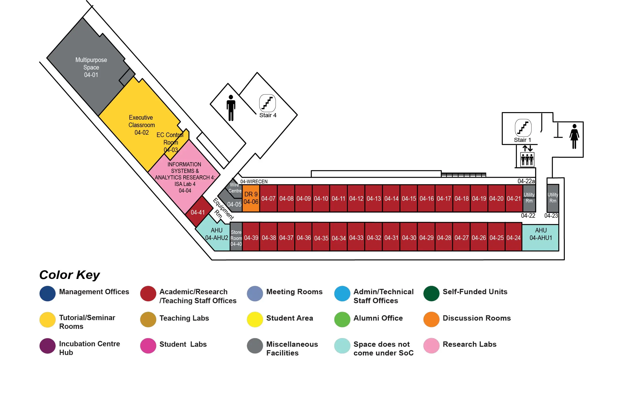
COM2 Level 4
Executive Classroom, COM2-04-02

Abstract:
Cancer is a group of diseases involving abnormal cell growth which may affect many body organs. General treatment of cancer involves assessment of cancerous tissues by the pathologist, followed by therapy prescribed by the oncologist according to the pathology report. Pathologists look for various cellular and cytological patterns while assessing the histopathological tissue slides. These patterns are heterogeneous and subjective. Eventually, the manual and tedious observation task impedes intra-observer agreement among pathologists.
In this thesis, various advanced algorithms for effective automated grading of cancers were developed. These algorithms enhance the pathologists' efficiency and reduce their errors. These algorithms can be integrated to give a more holistic assessment of cancers. These new algorithms focus on prostate cancer (PCa), breast cancer, and kidney cancer which are one of the leading causes of cancer deaths in Singapore.
Prominent nucleoli are important cytological patterns for renal cancer, breast cancer, and PCa assessment. Glandular architectures with their varying shapes and sizes are important patterns for PCa assessment. Cribriform pattern in glands is particular crucial for PCa assessment. Nuclear pleomorphic patterns along with prominent nucleoli are used for renal cancer grading. This thesis presents advanced algorithms for detecting these patterns in histopathological images to aid pathologists.
The analysis of prominent nucleoli is one of the several important considerations for cancer diagnosis. A prominent nucleoli detector was developed using various new feature extraction and classification methods. It is essentially a boosted combination of a cascade detector farm. As the detector can present most relevant information to the pathologist by its ranked nucleoli detections, it can reduce the manual task of the pathologist while going through large areas of whole slide images. Our detector performs twice as good as the use of a single cascade proposed in the seminal paper by Viola and Jones. The performance of our newly developed classification methods for the task of distinguishing between nuclei with and without nucleolus was also studied.
Renal cancer tissues typically contain cells with enlarged, irregularly-shaped and darkly-stained nuclei with prominent nucleoli. The prominent nucleoli detection system discussed above along with new feature extraction methods was used to develop an automated renal cancer grading system. The new feature extraction methods were designed to quantify the nuclear pleomorphic patterns and aid in distinguishing between renal histopathological images with different prognosis. We also demonstrate that the image score computed by our system is positively correlated with an existing multi-gene assay based scoring system which has been previously shown to be a strong indicator of a renal cancer patient's prognosis.
An automated gland segmentation system was developed to improve pathologists' accuracy and reduce their gland structure assessment time. This system uses machine learning methods for empirical segmentation and refines it further using image processing based methods. This system while outperforming various existing methods in the literature performed poorly in images with cribriform pattern due to its characteristic features. Cribriform pattern is a strong predictor for distant metastasis and disease-specific death. Subsequently, transfer learning coupled with deep learning was used to develop an automated cribriform pattern detection system.
This thesis focuses on the algorithm development for prominent nucleoli detection, automated renal cancer grading, gland segmentation, and cribriform pattern detection while aiming towards effective automated grading of cancers. These algorithms aid cancer care by enhancing the pathologists' efficiency and reducing their errors.

